The Benefits of Biological Therapies in Dentistry

Today homeopathy is an often used system of medicine which is not only safe, but also has no harmful side-effects. It involves remedies chosen on a highly individual basis. In the dental field it offers surprisingly effective therapies for toothaches, gum disease, abscesses, ulcers, children’s teething problems, dental fear and anxiety, etc. Since homeopathy acts on an energy level, it acts extremely well on children especially, because of their high energy, vitality and purity.

Herbal remedies can be used as teas which stimulate the body’s immune system and its detoxification mechanisms, and also as natural constituents in mouthwashes and toothpastes. Some herbs are not suitable for children, pregnant women, for those on anti-depressive or other drugs, or with liver damage. Please consult your physician before using herbs internally.

Aromatherapy, hypnosis, acupuncture, bioresonance, low level laser, biomeridian and bioenergetic therapy, applied kinesiology, osteopathic therapy and nutritional therapy can all have applications in dentistry. A biological dentist uses a multidisciplinary approach in utilizing these services, or referring a patient to specialists in complementary therapies, thus benefiting the patient as a whole.

Lasers in Dentistry

L.A. Health News, December 2004

The laser has now ultimately infiltrated the world of dentistry. This amazing technology, which has been the centerpiece of futuristic and science fiction myths, has actually found numerous applications in medicine over the past three decades. Nowadays, it is an everyday reality in an increasing number of dental offices. There are many types of dental lasers, but I will refer mainly to the ones that can be used on hard tissue such as teeth. Developed into streamlined machines and equipped with efficient delivery systems, these can be used to prepare cavities, sterilize root canals, treat periodontal pockets, trim gums, and assist in soft and hard tissue surgeries. Many of them work by emitting pulses of laser energy, which excite the water molecules on the surface of the target tissue, making them break away in microscopic layers. This is termed “hydrokinetic energy”, and since it does not involve heat and vibration, there is hardly any pain in most procedures.

Examples of their uses in dentistry are as follows:

  1. Preparing tooth cavities for placement of bonded fillings. Used in place of the drill, it gently removes tooth structure layer by layer, without even touching the tooth. Children love it. Used correctly, it is safer than the drill, in spite of misconceptions over the word “laser”. In fact, this piece of equipment is one of the safest gadgets used in the mouth. As the cavity is cleaned out, there is a simultaneous sterilization in effect, and a surface is created which produces a superior bond with bonding agents.
  2. Fissure sealants: It is a well-known fact that along with cosmetics, preventative procedures will lead the way in modern dentistry. Along with improved oral hygiene programs, cleaning out the deep grooves, or “fissures”, on the chewing surfaces of teeth, and placing bonded sealants in them, constitutes a significant measure in the prevention of dental cavities. The laser is ideal for this purpose, as it obliterates the soft, decalcified and decayed enamel, along with bacteria and organic matter – namely the precursors of cavities – found in the microscopic depths of these fissures.
  3. Preventing root canals: Most root canals arise after bacteria from deep cavities invade and infect the nerve chamber. Many times, however, the process of using a drill to clean out decay so close to a live, but compromised nerve, will irreversibly traumatize it through high-frequency vibrations, or even crush the dentine and drive bacteria deeper into the nerve. This by itself may be the last straw, and cause a root canal. The laser, on the other hand, causes no vibration, structural damage, or heat. It will gently remove infected tooth structure in close proximity to the nerve and kill bacteria, thereby greatly reducing chances of a root canal treatment.
  4. Debriding and disinfecting root canals prior to placement of the filler material. The laser beam unclogs the dentinal tubules and kills bacteria.
  5. Treatment of gum disease. Today there is irrefutable evidence that people with gum disease run a higher risk of heart disease and diabetes. Conventional deep cleanings of infected gum pockets often result in millions of bugs entering the bloodstream and causing havoc. The laser is used as part of a thorough gum treatment protocol to safely disinfect pockets, and stimulate the diseased epithelium before undertaking more invasive measures.

IV Anesthesia Information and Instruction

In-Office Anesthesia for Pediatric Dentistry
John A. Yagiela, DDS, PhD
Christine L. Quinn, DDS, MS

Advances in anesthesia techniques have made it possible to provide safe, reliable, and economical anesthesia services for children in the dental office. The following is a description of patients who might benefit from sedation or anesthesia, the types of anesthetic services provided in the dental office, the risks and costs of such services, and important instructions for the parent.

Children below the age of 5 are often unable to cooperate with needed dental care, especially if the treatment is extensive and time consuming. Children with previous bad experiences in the dental office or who have become frightened by scary stories told by siblings or other individuals also have difficulty receiving necessary care. A third group providing a distinct challenge to the dentist includes children with special medical or behavioral needs. In years past, options for treating such patients were limited. The child could be placed in a hospital and receive general anesthesia—an expensive, inconvenient, and time-consuming option. Alternatively, the patient could be treated by the dentist using physical restraints or sedative drugs to control behavior. Limitations to this approach include compromises in dental care, patient comfort, and either the effectiveness or safety of the sedation. Lastly, for the very young, treatment could be deferred until the patient is old enough to tolerate regular care. Problems with this approach include increased cavities and pain, and heightened risk of infection, premature loss of teeth, and malocclusion. In-office anesthesia provided by an independent dentist anesthesiologist offers a highly effective and safe solution to the child, parent, and dentist.

The dentist anesthesiologist is fully trained in outpatient anesthesia for the dental patient. He or she will evaluate the needs of the child—medical, dental, and psychological—and determine the most appropriate type of anesthesia service to provide. The dentist anesthesiologist will provide the necessary preoperative evaluation, intraoperative management, and postoperative care for the safe and effective treatment of the child. Additional responsibilities include ensuring that all necessary drugs, supplies, equipment, and facilities are immediately available and that the patient is continuously monitored throughout the case. The range of in-office anesthesia services available include specialized local anesthetic techniques, conscious sedation (in which the patient is relaxed but responsive to verbal command), deep sedation (in which the patient may not be responsive to verbal command), and light general anesthesia. Intubation general anesthesia can also be performed but requires additional drugs, supplies, and equipment.

The safety record of in-office anesthesia administered by an independent dentist anesthesiologist is unsurpassed by any other system of anesthesia delivery. Nevertheless, there are some potential complications that need mentioning. Nausea and vomiting, though uncommon with the forms of anesthesia delivered in the dental office, can occur. Generally, vomiting is most likely after the child wakes up and begins to move about. Venous irritation from the insertion of the intravenous catheter or from the drugs administered is another unlikely and minor potential complication. Because children vary in their response to medications, recovery may be delayed, especially after prolonged procedures. Laryngospasm, a condition in which the vocal cords close in response to a stimulus such as liquid dripping into the back of the throat, occasionally occurs and requires for its treatment suctioning of the mouth, delivery of oxygen under gentle pressure, and rarely the administration of a muscle relaxant. Respiratory depression, a possible side effect of all sedative agents, is easily managed by ensuring a patent airway and providing ventilation assistance. Allergic reactions, extremely rare with the drugs used in anesthesia for dentistry, are managed by the use of emergency drugs to reverse the signs and symptoms of the reaction. A final complication of anesthesia care in the child is urination during or after a prolonged procedure. A diaper or training pants is especially helpful in the young child, and bringing a change of clothes is advisable for all pediatric patients. A blanket is also useful in helping to ensure the patient is comfortable during and after treatment.

The following pre- and postoperative instructions are general in nature and may be modified by the dentist anesthesiologist to meet specific needs.

Preoperative instructions:

  1. Fasting: No solid food or milk for 8 hours before anesthesia. Children who are breast feeding may do so up to 4 hours before treatment. No clear liquids (e.g., water, apple juice) for 2 hours before anesthesia. Clear jello and popsicles are considered clear liquids.
  2. Medications: Take medications with a small sip of water.
  3. Clothing: The child should wear a short-sleeved shirt or blouse and nonrestrictive clothing. Small children should have on a diaper or training pants.
  4. Parents: A parent or guardian should be with the child and be able to give the child undivided attention (e.g., no small siblings with a single parent).

Postoperative instructions:

  1. Feeding: A clear liquid should be first offered to the child in small volumes. Soft foods (jello, ice cream, warm but not hot soup) may then be tried. Chewing should be avoided until all local anesthesia has dissipated.
  2. Temperature: A small elevation in temperature (under 101 °F) may occur the first day after anesthesia. It may be treated with children’s pain reliever. Avoid overdressing the child, especially on a warm day. If the child appears flushed, wet the skin with cool water.
  3. Sleeping: It is normal for a child to be sleepy after anesthesia. The child should be placed on a firm mattress without a large pillow.
  4. Call the dentist or dentist anesthesiologist if there is persistent vomiting (beyond 4 hours), if the temperature rises above 101 °F, or if there is any other concern.

UCLA Dental Anesthesia Service

Drs. John A. Yagiela and Christine L. Quinn are full-time faculty members of the UCLA School of Dentistry who provide in-office anesthesia services as a part of their faculty responsibilities. They are often accompanied by their second-year anesthesia resident to provide team care for the patient.

Dr. Yagiela received his DDS degree in 1971 from the UCLA School of Dentistry and his PhD in pharmacology in 1975 from the University of Utah. He received his anesthesia training at the UCLA Center for the Health Sciences and has a joint appointment as Professor of Anesthesiology in the School of Medicine, in addition to his primary appointment as Professor and Coordinator of Anesthesia and Pain Control at the School of Dentistry and Chair of the Division of Diagnostic and Surgical Sciences. A Diplomate of the American Dental Board of Anesthesiology, Dr. Yagiela has been providing in-office anesthesia services since 1984.

Dr. Quinn graduated with the DDS degree from the USC School of Dentistry in 1987 and obtained her MS degree and certificate in dental anesthesia in 1989 from The Ohio State University College of Dentistry. She is a Clinical Professor of Anesthesia in the School of Dentistry. A Diplomate of the American Dental Board of Anesthesiology, Dr. Quinn spends half her time in teaching and patient-care activities at UCLA and the remainder in delivering out-patient anesthesia services in dental offices.

(reprinted with permission)

Early Childhood Caries

Early Childhood Caries (ECC), or previously known as “bottle decay” is a phenomenon still poorly understood, but pretty widespread. Many parents notice their 2-4 year olds developing widespread cavities on their front teeth, in spite of the fact that they may be extremely cautious with their diet and oral hygiene. In the past it was associated with babies sucking on their formula milk bottle in bed and going to sleep with it, thus creating an environment for the decay-causing bacteria to thrive on the pooled and fermenting milk on the surface of their teeth. Nowadays there seem to be other aggravating factors, and thorough attention has to be given by parents to keep the teeth clean from the bacteria that cause this condition.

Diet, proper oral hygiene, Xylitol gels, some homeopathic remedies, and proactive dentistry can all be used to prevent or slow down this condition.

Early Childhood Caries Facts

    • ECC develops as a result of very early colonization of the baby’s mouth with streptococcus mutans bacteria.
    • The bacterial colonies covering the teeth, known as plaque, will process sugars coming into contact with them and produce acid as a by-product, which in turn will cause enamel to become softer and porous. These white chalky lesions will then progress into the more advanced decay seen in rotting teeth, especially in upper incisors of children between the ages of 2 and 4.
    • Even though recent accounts maintain that breastfeeding is not associated with a higher risk for early childhood caries (ECC), we see, however, in our practice a high incidence of ECC in breast-fed children ages 2-4, ironically children of very healthy and diet-conscious parents. This is especially pronounced in cases where nursing is carried out into and throughout the night. We therefore urge mothers to stop nighttime nursing and step up tooth cleaning methods using gauze and xylitol before the child goes to sleep.
  • Other independent associations with increased risk for early childhood caries in the literature are rather older child age, poverty, and maternal prenatal smoking. Some ethnic groups are also independently associated with severe early childhood caries. It is appropriate to state here that in primitive and indigenous cultures with primitive diets, the incidence of childhood and adult decay, periodontal disease and tooth crowding practically did not exist, according to the unequaled anthropological research done by Weston Price).

Dental Homeopathy

Homeopathy has definitely found its way into biological dentistry, and today many dentists are using varying homeopathic remedies for various purposes. The ailments of the mouth, including the teeth, are in essence small-scale versions of other bodily malfunctions, and sometimes even one of their many manifestations.

It is therefore understandable, especially in light of the heavy emotional aspects of dental diseases and their treatments, that homoeopathy would be a valuable method of supportive therapy.

The modern concept of biological therapy may include classical homoeopathy, complex homoeopathy, isopathy, homotoxicology, auto-sanguis therapy, Sanum (Enderlein) therapy, nosodes, etc.

Many of these have their origins in Germany, where homoeopathy is well integrated into and tolerated by mainstream medicine.

Homoeopathy can be used in dentistry for anxiety, pain, sensitivity after fillings, infections and abscesses, gum disease, before and after surgeries, dental trauma, ulcers, etc.

Constitutional treatment by a professional homoeopath will also use deeper acting remedies to treat chronic disease, strengthen an individual’s general health, emotional makeup and immune system.

To find out more about homoeopathy, go to ABC Homeopathy.

In A Dental Emergency

Chipped Teeth

Tooth trauma is very common in children due to their active nature. The upper front teeth are the main victims. The damage can be anything from a small chip to outright “extraction”.

A chip usually involves just the corner, small or large, half the tooth horizontally, or the whole tooth at the level of the gums. It creates panic due to its unsightly appearance. However, these teeth can be treated by a dentist experienced in bonding and aesthetic procedures.

A small chip: There is no urgency to see a dentist. However, a regular dental appointment should be made for a cosmetic bonding procedure of the chipped tooth.

Repair of a small tooth chip

A large chip involving dentine exposure: the tooth will be extremely sensitive to air, cold and hot drinks. Definitely visit the dentist within two days.

A large chip involving exposure of the nerve: If you see a small pink or bleeding point in the middle of the fracture area, you should visit the dentist as soon as possible. In the meantime, avoid noxious foods, cold and hot drinks, sweets, and even touching the area.

Chipped Front Tooth
Tooth Fracture Involving Dentine and Nerve Exposure
Repaired Chipped Front Tooth
After Laser Cap of the Nerve and Bonding with Composite

Restoration of a tooth fracture can be combined with the application of veneers, and the results can be amazing. This patient could not stop smiling after all was done.

You can see more pictures of this patient under Services – Veneers

Homeopathic remedies to consider: Traumeel and Hypericum 30c alternating once an hour on the day of injury.

Knocked Out Teeth

If your child falls and knocks out a tooth, do not panic. Find the tooth, pick it up carefully from the crown (not the root), rinse without rubbing the root, and preserve it in a saline solution such as Save-a-tooth®, contact lens solution or any eye-drop solution. Saliva can be used, milk is not recommended any more. Wrap it up carefully so it does not dry up. Take the child to a dentist as soon as possible. The tooth may actually be re-implanted and splinted for a few weeks. Giving homeopathic Traumeel or Arnica every half hour will help.

Children under 5: the tooth most commonly knocked out is an upper primary incisor. It is of no grave consequence if the tooth is lost, however esthetics and phonetics (development of speech and pronunciation) may be an issue.

Children between 6-8: The primary incisor is close to being shed and already loose due to the fact that the roots are much shorter. Do not bother re-implanting, as very soon the permanent incisor will appear.

Children 8 and older: Due to the fact that their new upper centrals are large and flared, and they are in a more physically active phase, this age group suffers the most incidents of knocked-out teeth. These teeth have to be preserved and reinserted at all costs, due to the following:

  1. The loss of these teeth have grave consequences on the child’s esthetic appearance and self-esteem;
  2. Definitive restoration of the lost tooth/teeth cannot happen until the age of 18, due to the child’s active jaw-growth phase. It will be limited to false teeth attached to active removable appliances that will allow for the natural growth pattern of the jaw. After 18, expensive bridgework or implants are the only choices;
  3. High and ongoing cost of providing interim and final restorations for these teeth.

It is evident that parents have to treat an incident like this more seriously than a bone fracture. A bone will heal and that’s the end of it. Unless it is kept alive and re-implanted, a tooth lost cannot be replaced, and will run expensive bills for years to come. If done early enough, the tooth will reattach, may however require root canal therapy in the future. It should be carefully monitored at every dental check-up appointment.

Dental Abscess

Abscesses commonly have two causes:

  1. Gum infection, or
  2. A dead nerve of the tooth.

In either case, the swelling and pain arises from the fact that the pus accumulating in the bone and tissue has no way to escape and causes an increase in pressure and pain. The body tries to defend itself by causing the blood vessels to swell and thereby directing white blood cells into the area to fight the infection. This battle releases breakdown products and more toxins which contribute to the pain and swelling.

If you experience such an infection visit your dentist immediately. If unable to get to a dentist, you should drink lots of fluids, vitamin C, and remedies that have a natural antibiotic effect, such as Echinacea, goldenseal, oregano extract, olive leaf extract, or homeopathic remedies which help in drainage and infection. Some single remedies useful in addressing abscesses are merc.sol 12c, hepar sulph. 12c, hekla lava 30c, graphites 30c, gunpowder 30c, among others.

Information for Parents Regarding the Oral Health of Their Children

It can be assumed that we are all aware of the major role we play in educating our children. In their search of guidance, they often look up to us and try to copy our example and our moral values. Issues like diet, health, as well as personal and oral hygiene are often related to an attitude which develops early in life and is profoundly influenced by the parents, who themselves are being bombarded nowadays by an endless flow of information and advertising, most of them contradictory. Responsible parents, therefore, should acquire a basic knowledge on health issues, if they are to protect, educate and raise their children along safe and unbiased guidelines.

When does our responsibility start regarding the teeth of our children?

When does our responsibility start regarding the teeth of our children?
BEFORE BIRTH! A healthy, stress-free pregnancy with adequate vitamins, minerals and organic foods will nourish the growing fetus and its tooth buds, which start calcifying within the jawbones already by the fourth month of pregnancy. This is also a good time to catch up on reading and preparing to take on new responsibilities. A word of caution: mercury circulating in the mother’s blood will actively accumulate in the fetus through the placenta. Therefore, during your pregnancy, and during the nursing period after giving birth, avoid any dental treatment, certainly those involving amalgam placement or replacement.

When is the right time to start brushing?

Preparations for brushing should start even before teeth erupt at 4-8 months. Rubber-tipped “toothbrushes” can be used to massage the gums to relieve the discomfort and itching associated with the teeth erupting, and to stimulate blood circulation. You should brush their teeth regularly as soon as they erupt. The baby will then realize that brushing is an important routine just like nappy changing. With a little imagination, games and stories may be associated with brushing to make it more fun.

By 3, a child should already manage simple brushing strokes on its own. As more teeth erupt, the parents should start flossing their teeth, in particular those teeth which are in tight contact .

How can we keep children away from decay – causing foods?

Never let your baby fall asleep with a bottle in the mouth. Milk, sweetened or acidic drinks will pool around the teeth and cause them to decay very fast. Children naturally have an instinct for eating simple, nutritious and vital foods. However, soon enough they are exposed to the colorful world of decay-causing sticky sweets and processed foods which give them more calories and less nutrients, all in a lifeless form. They are reprogrammed to demand more quantity and variety. Heavily sweetened sodas replace water, and constant, unhealthy snacks replace the nutritious meal. Today it is impossible to ban the child from having sweets and candy, but you can negotiate and compromise.

Hints:

Do not brush teeth right after sour or acidic drinks. Just rinse.

Nibbling on a piece of hard (non-creamy) cheese (preferably sheep or goat or tofu) will re-calcify the child’s teeth. Hard and chewy natural foods are important in stimulating the child to chew and produce saliva – along with the IgA antibodies present in it – as well as stimulate and massage the gums, and provide a self-cleansing action for the teeth.

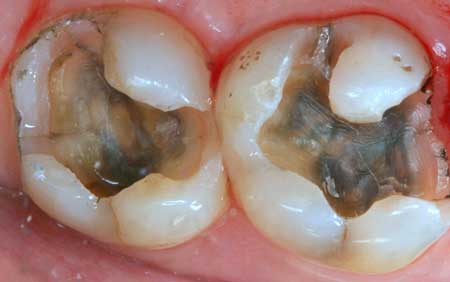
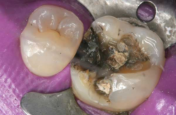
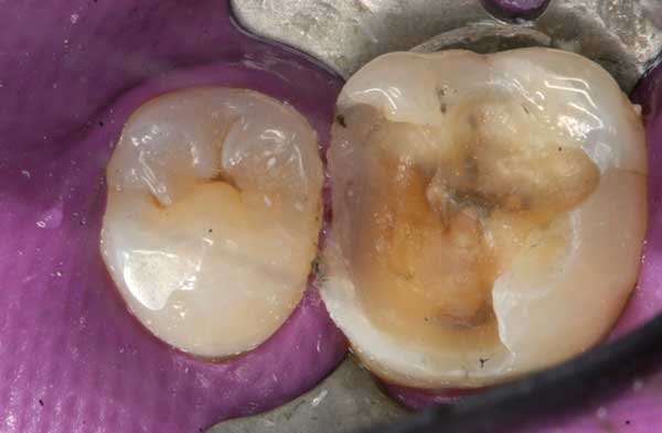
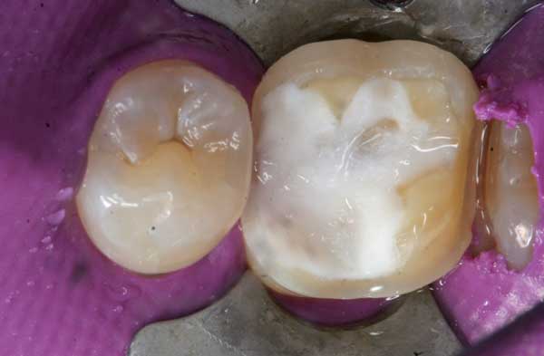
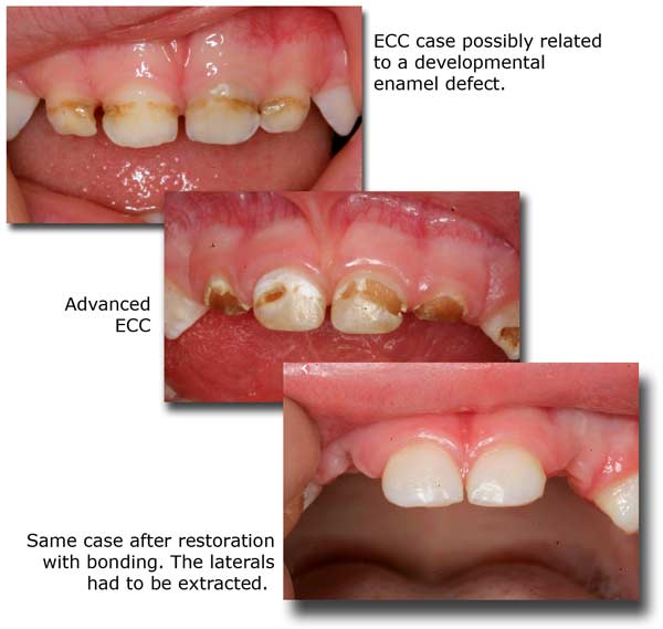
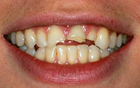
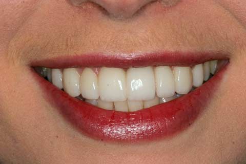
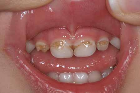
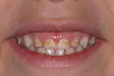
Since baby teeth will be replaced, is it all right to ignore cavities in them?

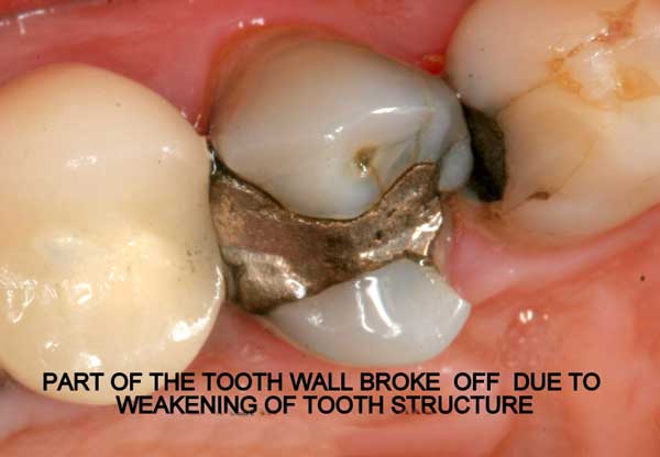
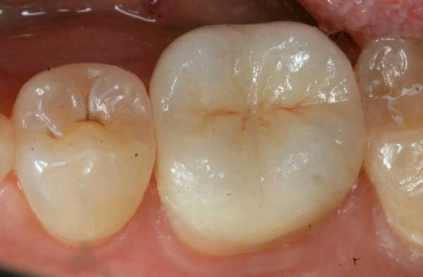
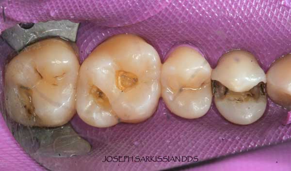
NO! Because of their smaller size and consistency, decay in baby teeth proceeds faster and reaches the nerve sooner than in permanent teeth. It is important to treat all cavities in these teeth because:

  • They are important spaceholders for the incoming permanent teeth and should be kept as healthy and as long as possible.
  • If they get abscessed, they not only cause unnecessary pain and psychological trauma, but they could also damage the underlying developing permanent tooth.
  • NOTE: LASER DENTISTRY is very appealing to kids and can be used to replace the drill and solve many fear issues. After all, it is the ultimate rewarding experience to see your child walk out of the dental office with a large smile and a feeling of positive accomplishment. Check out our website (sarkissiandds.com) to find out more.

When should we take the children to the dentist?

If you wait until the child has toothache, IT WILL BE TOO LATE! The treatment of a neglected cavity almost always involves some pain, and if it is associated with the first ever dental visit, it will create a deep psychological fear in the child. A child should accompany the parents to the dental office starting as early as one year of age to familiarize itself with the dentist, the office environment, and the dental instruments and procedures (as long as they are painless and in a relaxed atmosphere). Later, procedures such as polishing and filling small cavities will be tolerated and luckily the child will never develop a long-standing fear of dentists.
A child, like all adults, should visit the dentist at least every 6 months. However, they should be seen by a dentist immediately if:

  • They suffer a blow to the front teeth (even with no apparent harm).
  • Any extraordinary swelling, color change, or obscure pain appears within the mouth or on the face/neck area.
  • Permanent teeth fail to erupt for more than 2 months after shedding the baby teeth, or baby teeth fail to drop within 6 months of their time.

Is it OK if the child is “put to sleep” to receive treatment?

It is a great source of anxiety for any parent to see their child receive general anesthesia. It is best to avoid the situation by practicing preventative dentistry and catching all cavities early. However, some children under the age of 5, who have a predisposition to decay with multiple cavities involving the nerves, will benefit more from general anesthesia, for the following reasons:

  1. If properly carried out, the incidence of a life-threatening event during i.v. sedation is not so much higher than the administration of local anesthetics.
  2. The level of anesthesia is not so deep in dentistry. Breathing is not suppressed, and the child will come to within 5-10 minutes after completion of the procedure.
  3. The cumulative load of local anesthetics injected over countless appointments may turn out to be more toxic to the liver than a single load of I.v. anesthesia.
  4. The discomfort, fear and anxiety involved with such involved procedures including possible pulpotomies or root canals simply cannot be avoided.
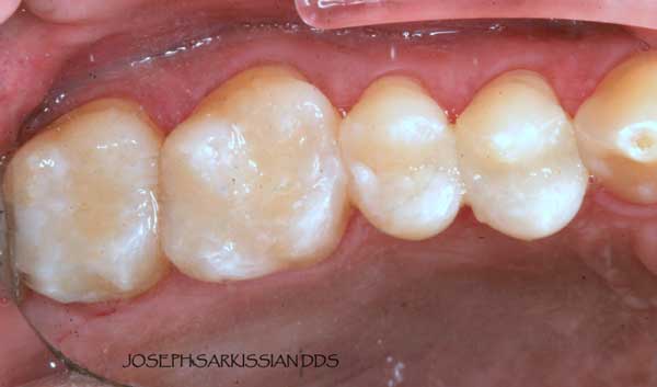
  5. The quality of work, particularly of such a nature as pulpotomies, bonded white restorations and metal-free primary tooth crowns can be significantly superior when performed under full anesthesia.

When is the time to consider orthodontics for a child?

A child’s face and skeleton follows a particular and complicated growth pattern, influenced by hereditary, environmental and functional factors. Any aberration of either of these factors will influence the growth pattern and will cause a disharmony. Many problems are a result of a discrepancy between tooth size and jaw size, which eventually lead to crowding. Allergies, deviated nose septums, hypertrophic adenoids and tonsils all cause mouth breathing, which start a cascade of skeletal facial anomalies such as retrognathic jaws, compressed dental arches and tongue thrust. Such growth patterns in turn lead to more serious airway deficiencies and sleep apnea. Other reasons are premature loss of a baby tooth, thumb-sucking habits, or tongue/lip dysfunctions including wrong swallowing patterns. Orthodontic evaluation and possible interceptive treatment of a child should begin at age 4. A sound orthodontic approach should always include influencing the natural craniofacial growth patterns with removable or functional appliances. Arch development using appliances at an early age, before the baby molars are replaced, is essential in your child having the necessary space to accommodate all the permanent teeth. Fixed braces and tooth extractions in many cases become unnecessary if this phase is successfully followed through. To find out more on appliance therapy, peruse this website.

Please see also Dr. Sarkissian’s website about Functional Jaw Orthopedics.

Important: Mouth breathing has the worst consequences. Address all causes (allergies, habits, adenoids) as soon as possible so lip closure and nose breathing resume.

Safe Amalgam Removal

ADA (American Dental Association) statement on amalgam:

Dental amalgam is considered a safe, affordable and durable material that has been used to restore the teeth of more than 100 million Americans. It contains a mixture of metals such as silver, copper and tin, in addition to mercury, which binds these components into a hard, stable and safe substance. Dental amalgam has been studied and reviewed extensively, and has established a record of safety and effectiveness.

Disclaimer:

The replacement of serviceable amalgams for health reasons by dentists is considered unethical by the ADA code of ethics. Dr. Sarkissian never convinces his patients to replace amalgam fillings unless there is evidence of failure or recurrent decay, or the patient prefers to replace them for cosmetic purposes. He will never guarantee any improvements in health conditions or diseases unrelated to the oral cavity, just on the basis of removing amalgam fillings.

Introduction

If you are one of millions who have educated themselves on the mercury amalgam controversy and have touched that subject on the web, we do not have to give you a long lecture. The purpose of this section is not to examine and debate its toxicity, nor to discuss methods of heavy metal detoxification. However, we feel inclined to provide a summary of the scientific facts about amalgam, its properties, its effects on teeth, and our precautionary protocols surrounding their removal.

Amalgam Ingredients

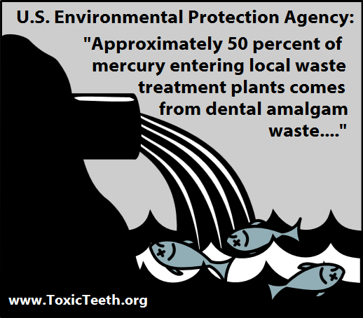
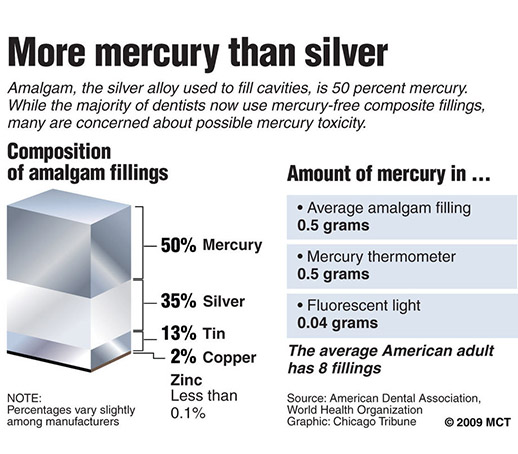
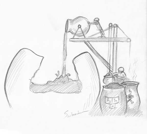
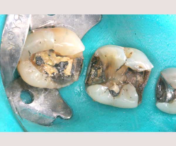
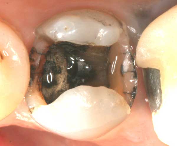
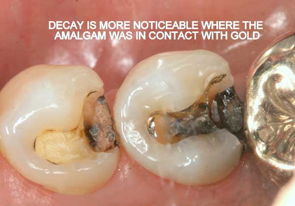
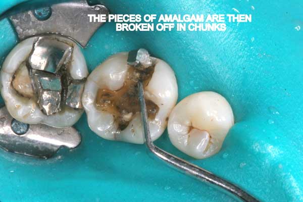
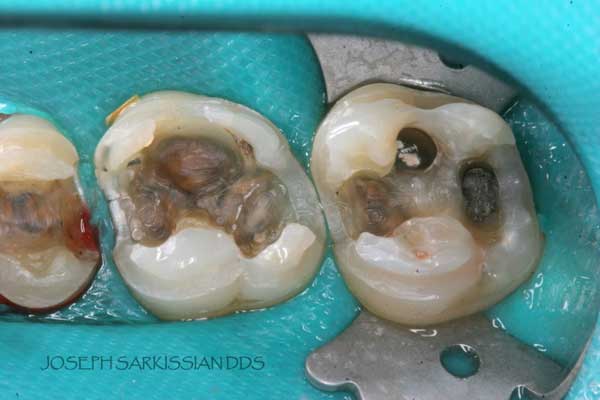
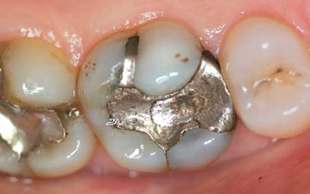
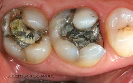
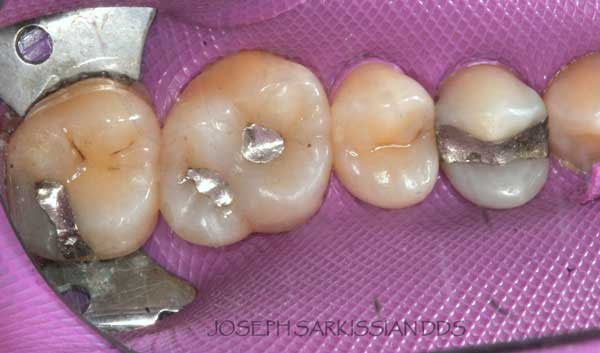
Half of what is known as amalgam is mercury, one of the most toxic substances known to man. The other half is made up of a powder of varying portions of silver, copper, zinc, and tin. As the liquid mercury and the powder are mixed, a paste is formed, which eventually hardens into an unstable “alloy”. In fact, it is not a true alloy, but a conglomerate of different “phases” between the mercury and the other metals, some of them somewhat stable, some less stable. Being unstable means that if the temperature of the amalgam “alloy” is raised, or its surface is agitated and/or rubbed, mercury vapor will be released in significant amounts. This does not happen with a true alloy.

History And Popularity

The main reasons amalgam caught on in the late 1800’s, (yes, you read it, more than 150 years ago) was the ease of placement, relative durability, and cost. Finally, there was a substance which would be affordable by almost anyone. After the initial furor and controversy surrounding its toxicity, it was eventually accepted and adopted by the ADA. Mercury amalgams were now being placed into the mouths of millions of people worldwide. Not to mention the vast amount of elemental mercury that was (and still is) entering the environment, in the form of human excrement, or as these metals are ground away by dentists, or as deceased amalgam carriers are being cremated.

Properties Of Amalgam As A Dental Restorative

Before a dentist places amalgam, the tooth cavity is first ground down into a particular form with undercuts. This unnatural gouging of the tooth weakens the cusps and creates internal strains. Second, the dentist packs the amalgam paste into the cavity and then carves it into shape before it hardens completely.

The following factors make amalgam an unsuitable material to use in teeth:

  1. Non-bondable nature promotes decay. There is no bond between the amalgam and the tooth structure. It is merely condensed tightly, and the only reason it does not fall out, is because of the retentive undercuts. The microscopic gap between the amalgam and the tooth will eventually allow for moisture and bacteria to creep in and cause recurrent decay. 99% of amalgams I replace harbor underlying decay. Most are completely undetected, until the damage is so extensive that the tooth will succumb to an expensive root canal treatment, or the walls will break off. In both cases, one faces extensive costs and discomfort.
  2. Expansion and contraction: Amalgam has significantly different physical characteristics than tooth structure. Being a metal, temperature fluctuations will cause it to expand and contract at a higher rate than the tooth structure. This discrepancy causes the already weakened tooth to crack and split. This constant “pumping” effect will cause cracks to propagate along the walls, under the cusps, sometimes even splitting teeth in half.
  3. Electrogalvanism: Amalgam is considered a non-noble metal which easily corrodes. This sets an electric current in motion, also called a galvanic current, which flows through the tissues, other metals, and the wet oral environment, to complete its circuit. The more dissimilar the metals, the higher are the currents. The worst currents are between gold and amalgam in direct contact.
  4. Amalgams are cosmetically unacceptable and ugly, particularly as we enter the 21st century. They are a violation of the esthetic, biomimetic and minimally invasive principles that modern dentistry promotes. Endowed with modern advances in technology and material science, today we have an abundant supply of acceptable, esthetically pleasing and durable options to restore teeth.
  5. Amalgams are a significant source of mercury exposure: It is a patient’s personal option to have amalgam fillings placed, or prefers to replace them. There is no direct link between amalgams and a particular disease, but there is ample evidence that mercury from amalgams is released, and that this mercury can be stored and traced in our organs. The fact that tooth grinding or exposure to higher temperatures causes an accelerated release of mercury vapor from amalgams is not something that one can brush aside. There is evidence that the amount of mercury stored in some organs is in direct proportion to the number of amalgams in a person’s mouth. In many countries the placement of amalgams in children, in pregnant women, or in those who have kidney impairment, is discouraged.

Safe Amalgam Removal Protocols

The main issue surrounding replacement of mercury fillings is to minimize mercury release into the surrounding air and environment, and to prevent exposure to patient and staff alike. This rule should be followed by ANY dentist replacing amalgams, not only biological dentists. It is unavoidable that some mercury is released when drilling out these old fillings, but with the right precautions the risk of ingesting this poison can be eliminated for the patient as well for the dentist and his assistant.

  1. Rubber dam: A rubber sheet is stretched around the tooth to isolate it from the rest of the patient’s mouth.
  2. “Clean-up” suction tips (IAOMT approved) are used as an alternative in case rubber dam cannot be applied. (image from the IAOMT web site)
  3. Slow speed drill and thorough water cooling is absolutely essential in reducing mercury vapor emission. We use a high-torque electric drill at 40,000 RPM for this purpose, as opposed to the 400,000 RPM turbine drills.
  4. Chunk removal: Amalgam is sectioned and then manually removed in chunks. This is possible since amalgam is not bonded to tooth structure. This way the actual drilling of amalgam into fine particles, as well as mercury vapor release is minimized.
  5. Dent-air-vac, a high-volume air filtration system with three layers of filters is used to clean the air in the immediate vicinity of the patient’s head during drilling. One of the filters inside is an activated charcoal filter which absorbs mercury vapor before the air is re-circulated.
  6. Oxygen administration via a nasal cannula, which in turn is covered with a moist napkin, ensures that the patient is breathing in clean oxygen during the drilling process.
  7. Activated charcoal in suspension is given to the patient to drink just before the procedure, in order to absorb any mercury that escapes despite the precautions.

Most small amalgams with moderate decay and no cracks can be restored with composites. These are BPA-free tooth-colored materials which are made up of a resin-based matrix filled with microscopic granules (nanoparticles) of ceramic and glass.

Larger cavities or teeth which have cracks are always restored with bonded porcelain onlays, which are very durable and biocompatible. The porcelain covers the cusps, protects the walls from further damage, and withstands chewing pressures. Their design is less invasive than full crowns, as healthy tooth walls are not ground down.

Showcases

Following are a few showcases of Dr. Sarkissian’s own patients, outlining amalgam replacement, the damage caused to the teeth, and how they look like after being restored with a biocompatible substance (composite or onlay).

Showcase 1: Amalgam revision with porcelain onlay

Showcase 2: Amalgam revision with porcelain onlay

Showcase 3: Amalgam revision with porcelain onlay

Showcase 4: Amalgam revision with composite

Showcase 5: Amalgam revision with composite

Links and References

Recommendations prior to Amalgam Removal

Please follow these recommendation for supplements to be taken and guidelines to be followed before and during amalgam removal. It must be stressed that they are based on varying nutritional protocols reported in complimentary medical and dental literature, and are not meant to treat any systemic diseases, nor to prevent any dental complications that may otherwise occur.

WARNING: We urge you to consult with your physician or nutritionist before taking any of these supplements , especially if your health or excretion is compromised. Please be aware that some herbs interfere with allopathic medication, or may be absolutely contraindicated for use in pregnancy, heart disease or antidepressant therapy.

  • Zinc gluconate or picolinate: 25-30mg 1/2 hour before bedtime.
  • Selenium: 200mcg once daily after breakfast. Do not take at the same time as zinc.
  • Calcium/Magnesium: 1000mg/300mg once daily after a meal. Taurate or orotate forms preferred.
  • Vitamin C: At least 2000mg twice daily after meals. Combine with bioflavonoids.
  • A liver herbal detox remedy supported by a homeopathic liver/gall bladder remedy. A “liver flush” is also advisable.
  • A homeopathic lymph support remedy.
  • A kidney herbal/homeopathic remedy.
  • A colon stimulating supplement including psyllium seeds and other fibers.
  • 3 capsules of activated charcoal twice a week in-between meals (take at least 1 hour away from all herbs and medications).
  • A chlorella/spirulina greens supplement.
  • Probiotics – intestinal recolonization with beneficial bacteria.
  • N-Acetylcysteine, 500mg dialy.
    Garlic – freeze-dried or high-grade extract.
  • Cilantro extract
  • Homeopathics individualized for detox
  • Also recommended are glutathione, Vitamin E and Omega fatty acids.

It is generally recommended to be on these supplements starting at least 2 weeks before any dental treatment and continuing for one more month after completion of therapy.

Throughout the weeks or months of treatment, it is advisable that:

  • A diet rich with protein and vegetables, but low on carbohydrates be followed.
  • No fish (including shellfish) be consumed. Sardines or other small fish are OK.
  • No chewing gum.
  • Bowel movements of 1-2 times daily be encouraged.
  • Plenty of liquids be consumed.
  • Sweating be encouraged either by exercise and/or saunas.

Biological & Homeopathic Dentistry

L.A. Health News, September 2004

Please realize that biological dentistry is not a true specialty in dentistry, but rather an approach to dentistry which utilizes some aspects of complementary (or “alternative”) therapy.

A biological dentist:
  • Will evaluate a patient as a whole person rather than a set of teeth.
  • Will respect the intricate inter-relationship between the teeth, the mouth and the body.
  • Will definitely not place any silver-mercury amalgams, and if he has to remove any, will follow strict precautionary guidelines.

Beyond these common points, biological dentists will differ in their methods. Some are trained in acupuncture, some in surgeries, bone infections or joint diseases, some in homeopathy, some in hypnosis. Overall, biocompatibility issues are of prime concern to them.

MOUTH/BODY CONNECTION

Diseases of the teeth and the mouth are in essence small-scale versions of other bodily ailments. Many times a disease will manifest itself first in the mouth. Recent studies indicate a strong relationship between bacteria involved in gum disease, and diabetes, coronary heart disease, and joint and kidney inflammation. Bacterial toxins from chronic bone infections or failed root canals, sometimes called “focal infections”, most of which remain unnoticed by the patient, have even more serious and debilitating health consequences.

In this context, doctors in Switzerland and Germany have established a detailed tooth/organ relationship chart to act as a guide for various practitioners.

TEETH AND EMOTIONS

The teeth, being in a part of the body essential to primitive survival instincts such as feeding, fighting, and facial expressions, are linked to deeply emotional centers in the brain. Many times dental treatment will trigger old memories and arouse long-forgotten emotions.

Granted, there will always be some sort of displeasure in going to the dentist. Nowadays, there are many ways to make dental treatment more acceptable. A serene setting, an understanding, open-minded dentist, and plenty of dialogue. Herbal relaxants, aromatherapy, acupressure, Bach flower remedies, homeopathic remedies for anxiety, calming music, distracting movies, hypnosis, acupuncture, all contribute to a serene and positive frame of mind and an optimistic attitude, thus creating conditions for speedier healing, and a better and more successful outcome in therapy.

HOMEOPATHIC DENTISTRY

Many biological dentists use homoeopathic remedies for various purposes. Homeopathy has been used for centuries and relies on the principle of “like curing like.” The remedies usually consist of extremely high dilutions of a substance, where there are no molecules left in solution, but rather the energy “imprint” of that substance on a submolecular level. This will resonate through the body and create a healing effect.

Depending on the dilution, or potency, a remedy can have differing depths of action, ranging from the biochemical/cellular level, down to the emotional/spiritual. In light of the heavy emotional aspects of dental diseases and their treatments, and considering the burden of heavy metal toxicity plaguing more and more people, homoeopathy could be a valuable method of supportive therapy. The broader concept of modem homeopathic therapy may encompass classical or complex homeopathy, homotoxicology, auto-sanguis therapy, nosodes, etc.

Many of these have their origins in Germany, where homoeopathy is well integrated into and tolerated by mainstream medicine. Overall, these modalities can be used in dentistry to address anxiety, pain, sensitivity after fillings, infections and abscesses, gum disease, healing after surgeries, dental trauma, ulcers, and detoxification.